03Nov
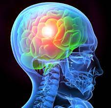
The Brain stem
The brainstem is the pathway for all fiber tracts passing up and down from peripheral nerves and spinal cord to the highest parts of the brainLocation: The brainstem is located at the juncture of the cerebrum and the spinal column.
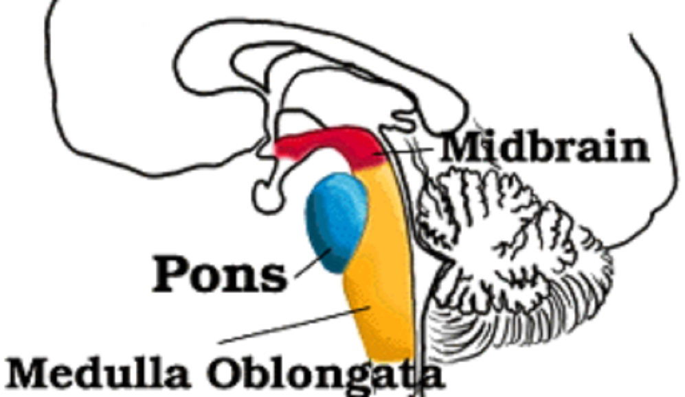
It consists of the midbrain, medulla oblongata, and the pons. Medulla Oblongata.
⦁ Respiratory Center :rhythmically stimulate the intercostals muscles and diaphragm — making breathing possible
⦁ Cardiac Center :regulate heartbeat .
⦁ Vasomotor (nerves having muscular control of the blood vessel walls) Center: Regulate the diameter of arterioles thus adjusting blood flow.
⦁ Centers for cough, gag, swallow, and vomitAssociated Signs and SymptomsPupils:
⦁ Size: Dilated.
⦁ Reactivity: FixedLOC: Comatose.
Respiratory:
⦁ Abnormal breathing patterns.
⦁ Ataxic. ⦁ Clustered.
⦁ Hiccups.
CN Palsies (Inability to control movement):
⦁ Absent Cough.
⦁ Gag.
Pons
⦁ Nerve impulses coming from the eyes, ears, and touch receptors are sent on the Cerebellum.
⦁ The Pons also participates in the reflexes that regulate breathing.
Associated signs and symptomsPupils: - Size: Pinpoint LOC:
⦁ Semi-coma
Movement:
⦁ Abnormal extensor.
⦁ Withdrawal.
Midbrain
⦁ Nerve pathway of cerebral hemispheres
⦁ Auditory and Visual reflex centers
Cranial Nerves:
⦁ CN III - Oculomotor (Related to eye movement), [motor].
⦁ CN IV - Trochlear (Superior oblique muscle of the eye which rotates the eye down and out), [motor].
Associated signs and symptomsPupils:
⦁ Size: Midposition to dilated.
⦁ Reactivity: Sluggish to fixed.
LOC (Loss of consciousness): Varies
⦁ Movement: Abnormal extensor (muscle that extends a part).
⦁ Respiratory: Hyperventilating.
⦁ CN (Cranial Nerve) Deficits: CN III, CN IV.
FORCES OF IMPACT
• Acceleration - caused by rapid increase in speed of head when impacted by moving object
• Deceleration - occurs when head stops suddenly
• Coup injury - brain injury directly below site of impact
• Contacoup injury - brain injury in region directly opposite site of impact.
Traumatic brain injury (TBI) is a nondegenerative, noncongenital insult to the brain from an external mechanical force, possibly leading to permanent or temporary impairment of cognitive, physical, and psychosocial functions, with an associated diminished or altered state of consciousness.
Types of brain injuries:
(1) Primary injury, which occurs at the moment of trauma
(2) Secondary injury, which occurs immediately after trauma and produces effects that may continue for a long time.
Types of primary injuries
Primary injuries can manifest as focal injuries (e.g., skull fractures, intracranial hemorrhages -hematomas, lacerations, contusions, penetrating wounds, concussion), or they can be diffuse (as in diffuse axonal injury).
Concussions: mild damage to brain characterized by brief loss of consciousness; impact mildly jostles brain inside skull; damage to gray matter of cerebral cortex, Diencephalon, or brain stem
Secondary types of traumatic brain injury (TBI) are attributed to further cellular damage from the effects of primary injuries. Secondary injuries may develop over a period of hours or days following the initial traumatic assault.
Causes of secondary injury
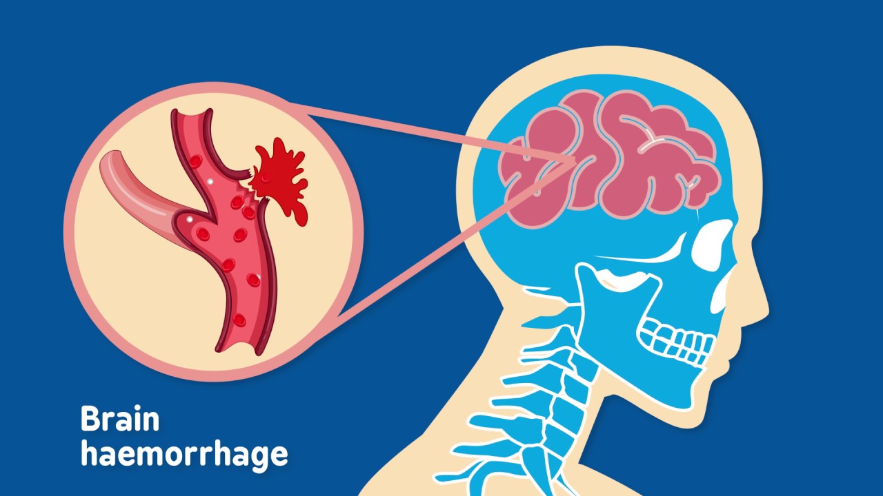
All tissues in the body swell when traumatized.The more damage the brain receives, the more it swells.
This is called brain swelling and occurs when there is an increase in the amount of blood to the brain. Later in the illness, water may collect in the brain which is called Brain Edema.
The Kellie-Monro Doctrine describes that the volume within the skull is fixed, and that an exponential increase in pressure occurs secondary to processes such as hemorrhage or swelling add to that volume. Such pressure elevation is called Increased Intracranial Pressure; it can impair blood flow to the brain.
Video Courtesy: Brain & Spine Foundation
TYPES OF HEAD INJURIES
Diagnosis
⦁ CT (computed tomography, uses x-rays and injection of an intravenous dye to visualize the lesion)
⦁ MRI (magnetic resonance imaging, uses magnetic fields and radio waves to visualize a lesion)
⦁ 1. radiological
⦁ * Skull x-ray
⦁ Cervical spinal x-ray (rules out cervical fracture)
⦁ ABG